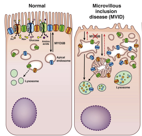
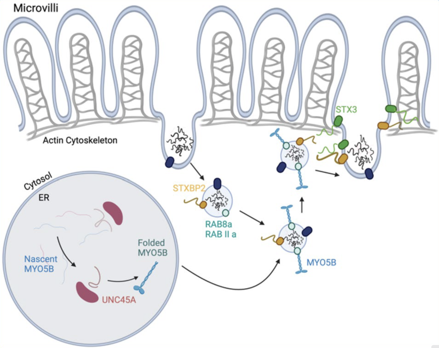
Microvillus Inclusion Disease (MVID) is a rare disease of the digestive tract characterized by an alteration of the microvilli of the small intestine, resulting in profuse and intractable watery diarrhea in children from the first days of life, leading to dehydration, malabsorption and growth retardation.
MVID is one of the so-called congenital enteropathies.
It is a genetic disease with autosomal recessive transmission.
Prevalence is estimated at around 200 patients diagnosed in Europe, a figure that could rise with better knowledge of the disease and diagnostic tools.
Clinical presentation:
Infants with MVID usually present symptoms soon after birth. Common symptoms include vomiting, severe watery diarrhea that is profuse and intractable, leading to severe dehydration and stunted growth.
Stool production is 100 to 500 mL/kg/day when the infant is fed, a volume comparable to or greater than that observed in cholera. Diarrhea is of the secretory type, and therefore persists even when fasting.
Due to high digestive losses, patients can lose up to 30% of their body weight in 24 hours, resulting in profound metabolic acidosis and severe dehydration.
Infants rapidly become dehydrated unless vigorous intravenous rehydration is started.
Complications
Complications of MVID are mainly due to malabsorption of nutrients and loss of fluids through diarrhea:
Dehydration: Frequent diarrhea leads to severe dehydration, which can be dangerous, especially for infants.
Malnutrition: Because of their difficulty in absorbing nutrients, people with MVID suffer from severe malnutrition, which affects their growth and development.
Growth retardation: Infants affected by this disease cannot gain weight properly and do not develop normally.
Medical complications: Infections and electrolyte disorders are increased risks due to the child's fragility.
Treatment
To date, there is no cure for the disease. Management involves total parenteral nutrition for life (administration of daily nutritional intake by vein), intestinal transplantation (when parenteral nutrition is no longer possible in the long term), and, if necessary, associated liver transplantation.
Current therapeutic options are limited, treat only symptoms, and have a significant impact on patients' (and caregivers/parents') quality of life, mainly linked to total parenteral nutrition.
The diagnosis is suspected on the basis of clinical manifestations, and is confirmed by histological analysis of small intestine biopsies revealing villous atrophy and abnormal inclusion material positive to PAS (Schiff's periodic acid) staining in the intestinal epithelium, without cryptic hyperplasia. Electron microscopy reveals microvillous atrophy, and in most cases, microvillous inclusion vesicles in the cytoplasm of enterocytes. Molecular genetic analysis has become essential to confirm the diagnosis.
The genes known to date to be responsible for the disease are Myo5b, STX3, STXBP2 (Munc18-2) and UNC45A.
Differential diagnosis(es)
The differential diagnosis includes rare congenital enteropathies, such as autoimmune enteropathy, chloride diarrhea, congenital sodium diarrhea and congenital tufted enteropathy.
Prenatal diagnosis
MVID is a rare disease with no systematic prenatal manifestation and no specific signs. Prenatal diagnosis is only possible if the specific genetic mutation has been identified in an affected family member.
Pathophysiology
The disease is linked to a loss-of-function mutation in one of the 4 genes known to date (stop, non-sense or mis-sense mutation):
Myo5B
STXBP2 (or Munc18-2)
STX3
UNC45A.
Mutations result in a polarization defect in the enterocyte and an accumulation of intracellular vesicles at the apical level (impaired exocytosis). The main transporters and ion channels are therefore not present at the surface of the intestine, resulting in defective absorption and significant losses that can be fatal if not treated rapidly.
Illustration of a breast enterocyte (left) and a mutated enterocyte typical of MVID (right)3
Apical surface of healthy (left) and mutated (right) enterocytes, scanning electron microscopy4
MVID is an endosomal trafficking disorder characterized by defective recycling of the apical membrane.
In healthy intestinal epithelium, early endosomes sort cargo vesicles into three main pathways: the retrograde Golgi transport pathway, the degradation pathway and the recycling pathway. The apical recycling pathway in intestinal epithelial cells is responsible for returning cargo vesicles to the cell membrane. It relies on the motor activity of Myosin5B along the actin cytoskeleton. Rab8a and Rab11a act as links between the recycling endosome and the C-terminal cargo-binding domain of myosin Vb.
Once the cargo vesicle has been delivered close to the cell membrane, interaction between syntaxin-3 t-SNARE (STX3) and the Sec/Munc-2 family syntaxin-binding protein (STXBP2, also known as Munc18-2) is required for membrane fusion.
Together, these proteins form a Myo5b-STX3-STXBP2 axis required for proper apical recycling, and loss of any of these major components results in an intestinal MVID phenotype. Although Rab8a and Rab11a are also vital for this pathway, there are no MVID-associated variants in either of these proteins.
UNC45A, a chaperone that assists Myo5b folding, has also become recognized as a prerequisite for proper apical recycling .
It is the combination of loss of epithelial surface area, loss of epithelial transporters, loss of appropriate vesicular trafficking and permeable tight junctions that is ultimately responsible for malabsorption in these patients.
For more information on the pathophysiology of MVID, please consult the Research page.
Ruemmele, F. M., Schmitz, J. & Goulet, O. Microvillous inclusion disease (microvillous atrophy). Orphanet J. Rare Dis. 1, 22 (2006).
Abtahi, S. & Turner, J. R. Exploiting alternative brush border trafficking routes to treat microvillous inclusion disease. Gastroenterology vol. 159 1233-1235 (2020).
Vogel, G. F. et al. Disrupted apical exocytosis of cargo vesicles causes enteropathy in FHL5 patients with Munc18-2 mutations. JCI Insight 2, (2017).
Babcock, S. J., Flores-Marin, D. & Thiagarajah, J. R. The genetics of monogenic intestinal epithelial disorders. Hum. Genet (2022) doi:10.1007/s00439-022-02501-5.